Corneal Flap Lasik
Crstoday Persistent Lasik Flap Interface Fluid After Dsaek Procedure
/flap_edge(425).jpg)
Lasik Flap Dislocation The Flap Never Heals

Corrective Eye Surgery 1 Ziyi Kuek Skat3rgalz

6 Common Questions About Lasik Eye Surgery Penn Medicine
Intraoperative Flap Complications In Lasik Prevention And Management Of Free Flaps Springerlink

A Clear Look At The Lasik Flap
Conclusions Acute corneal hydrops is a rare complication of postLASIK corneal ectasia In the absence of flap dehiscence and wound leak, such patients may be managed with simple observation.
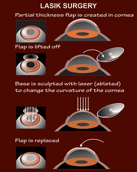
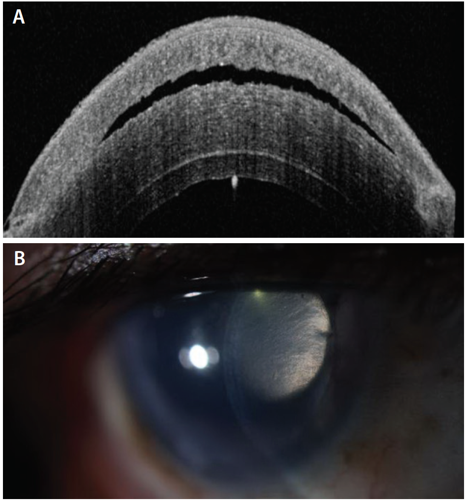
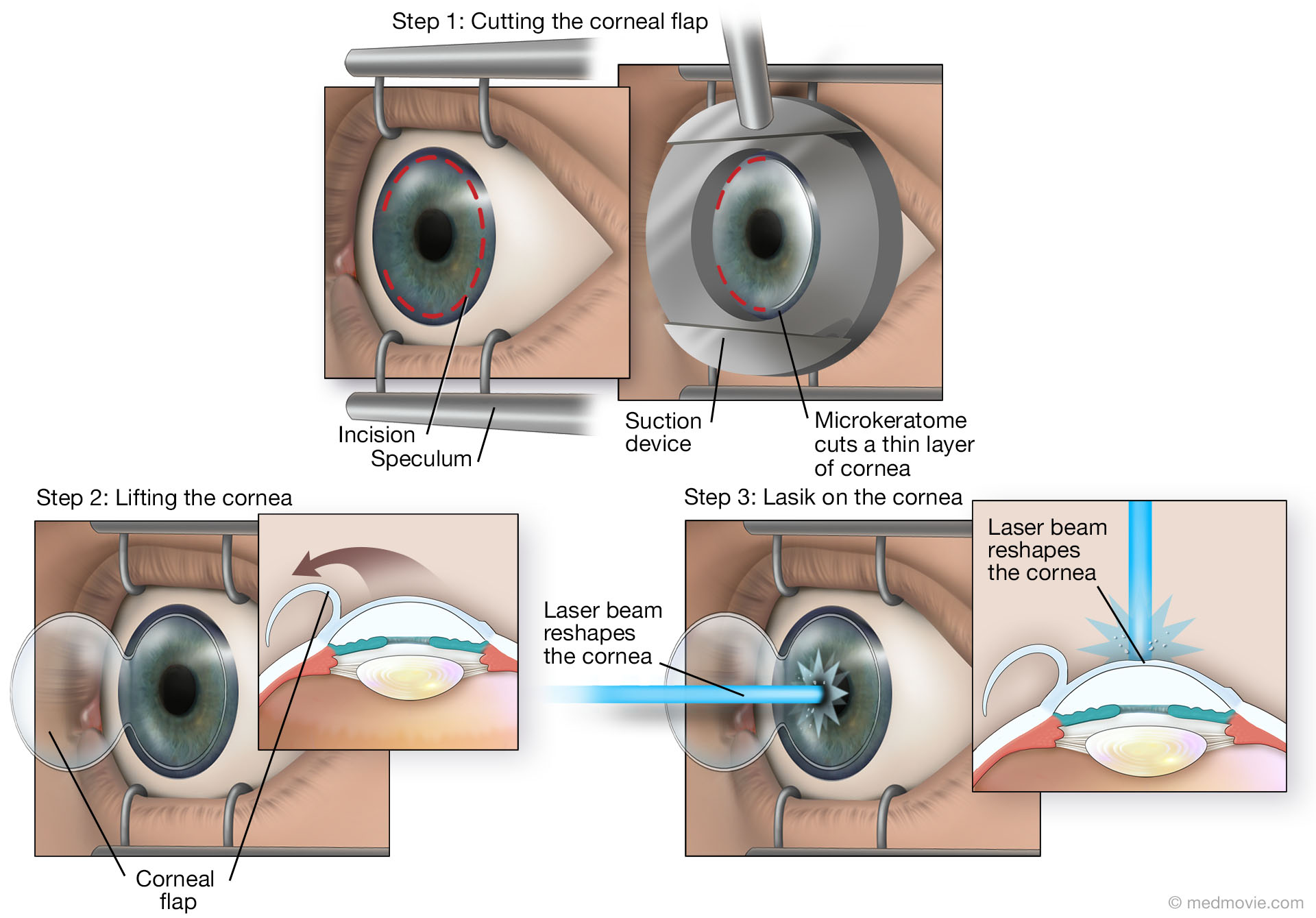
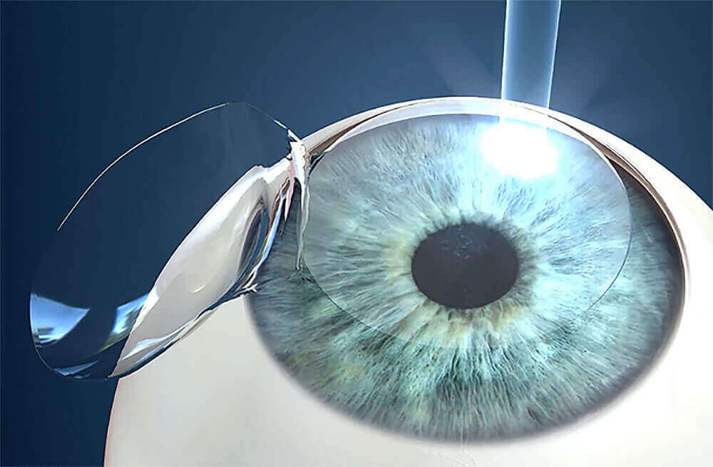
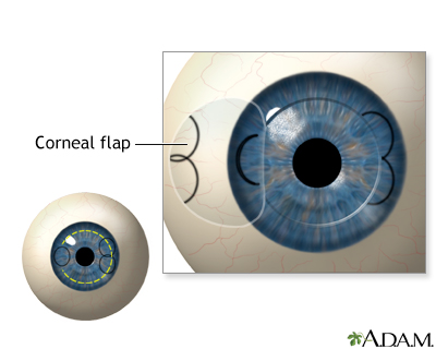
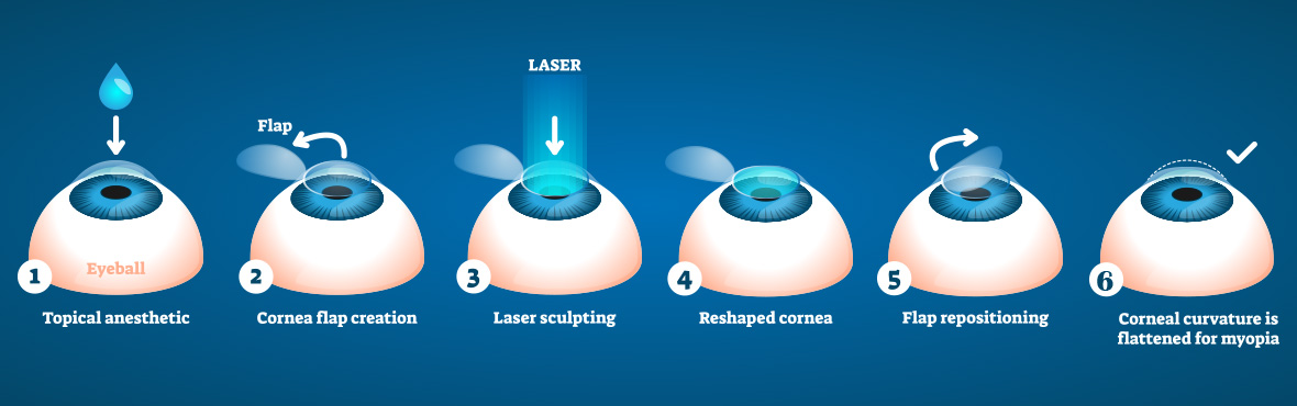
Corneal flap lasik. The next photo shows a cornea with two LASIK flaps Notice two arcs extending from 6 o'clock to 2 o'clock The patient had a second LASIK procedure several weeks after primary LASIK, and the surgeon cut a second flap within the original flap rather than lifting the flap Below is a cross sectional image of the cornea of a postLASIK patient. The corneal flap begins healing immediately after the LASIK procedure In fact, with the use of a LASIK flap, the corneal tissue can be as much as 90% healed within 24 hours During the first day or two after surgery, the outer surface of the cornea, known as the epithelium, seals the edges of the corneal flap. LASIK is a twostep process in which the cornea is first prepared for treatment by creating a corneal “flap” This flap is then utilized to temporarily expose a deeper layer of the cornea that a laser can precisely reshape to give the ideal corneal curvature to match a patient’s glasses or contacts prescription.
When performing all laser LASIK, our physicians use a gentle laser to create the corneal flap This technology, also used to facilitate laser cataract surgery, allows them to better customize the corneal flap for each patientAs the name states, no blades are used in this kind of laser eye surgery. Flap striae are defined as small wrinkles or folds in the cornea as a result of LASIK surgery Classification The most common classification of flap striae divides them broadly based on their size Microstriae are microscopic, superficial wrinkles that are generally asymptomatic and often only detectable on microscopic examination of the cornea. The creation of a flap in the outermost layer of the cornea is what distinguishes LASIK from other forms of laser refractive surgery, such as PRK and LASEK This means that flap complications are a potential risk of all forms of LASIK.
LASIK is a treatment for the correction of myopia, hyperopia and astigmatism It has advantages over photorefractive keratectomy, such as less postoperative pain and more rapid recovery, but. LASIK works by removing (ablating) tissue from the cornea This reshapes the surface of the cornea to correct the refractive error The amount of corneal tissue that is removed from the stroma is call the ablation depth, and this depends on the degree of refractive error. LASIK creates a thin flap in the cornea PRK removes the outer layer of the cornea, which grows back over time Both procedures have pros and cons Ask your doctor which one LASIK or PRK is.
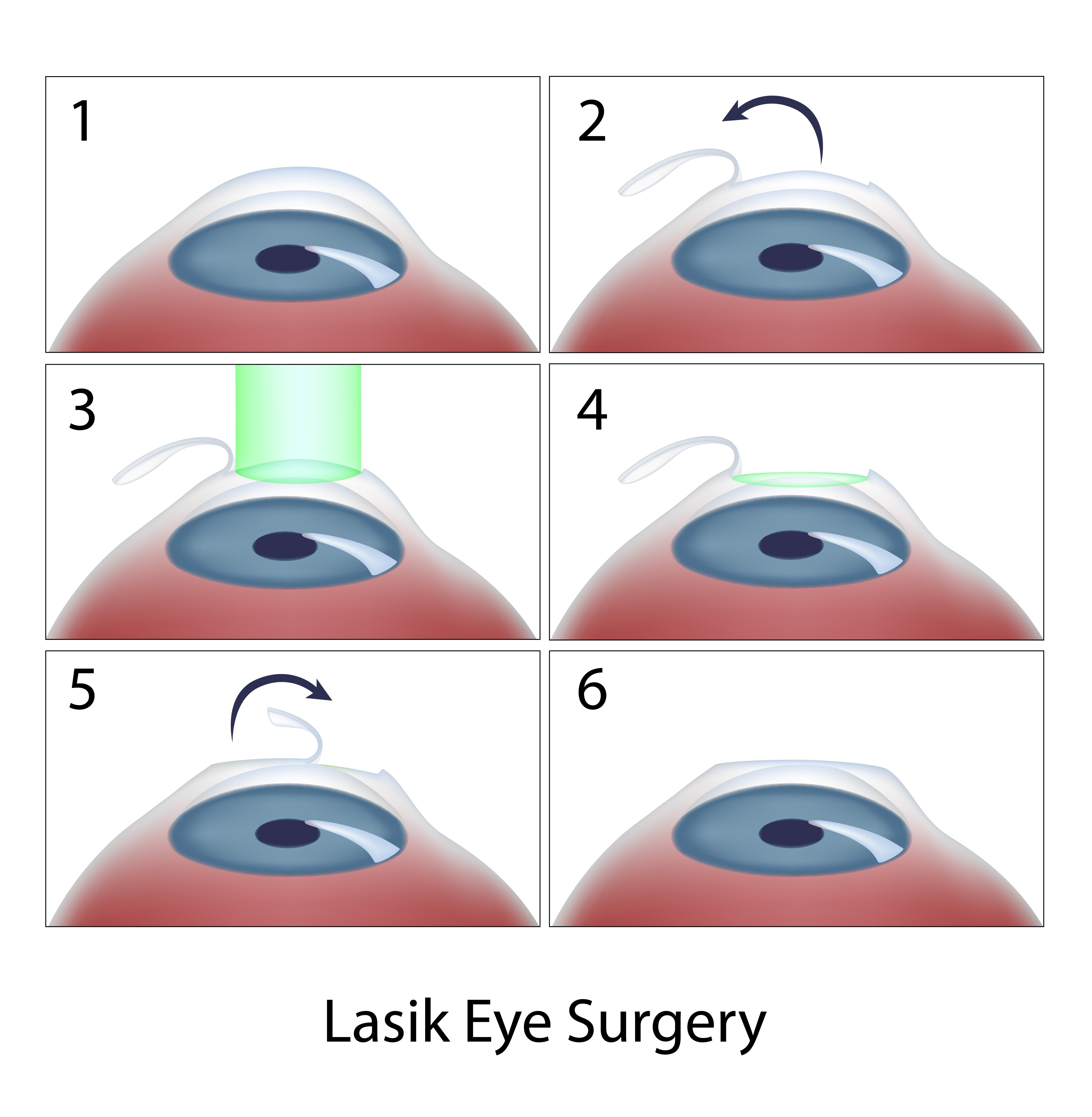
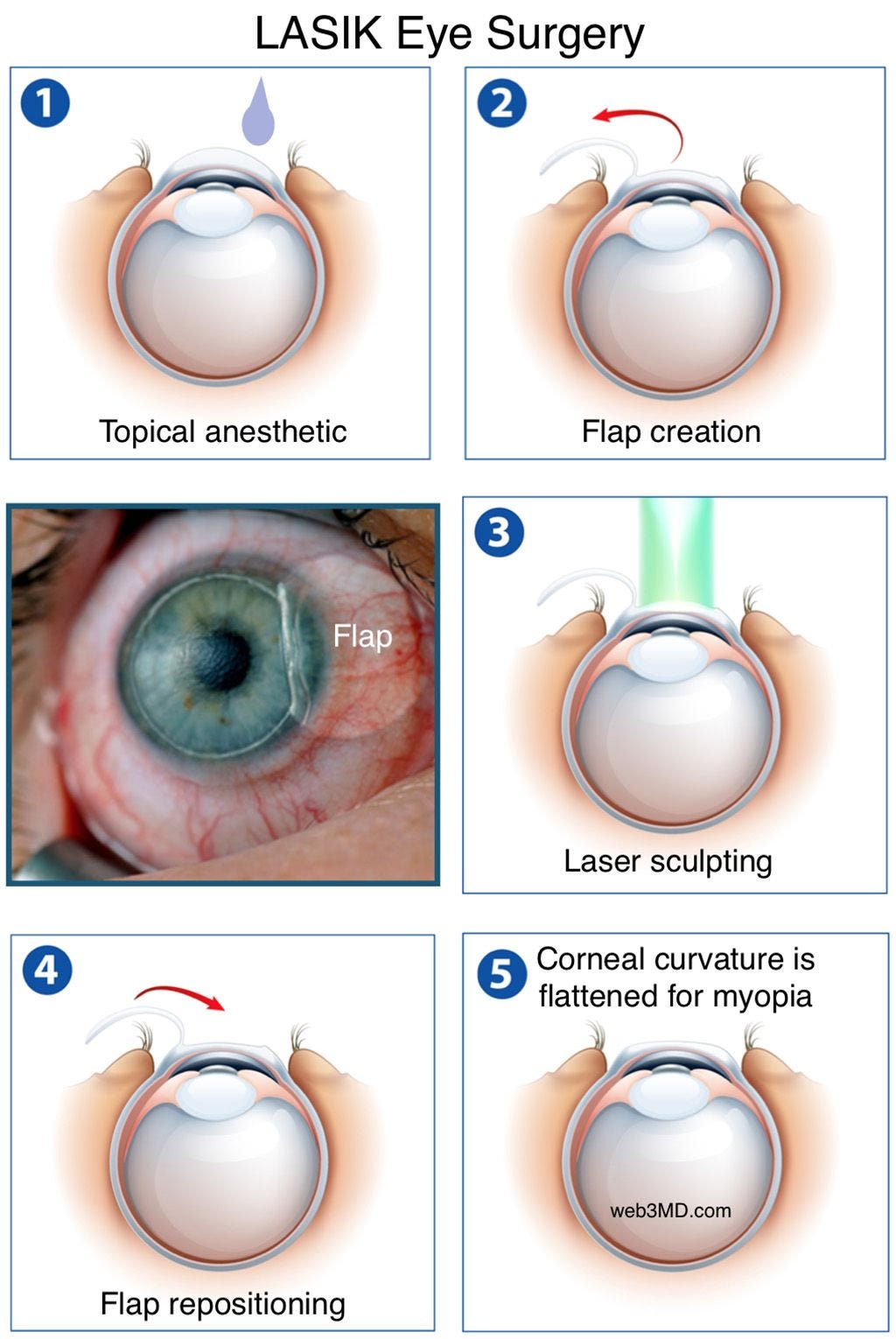
Corneal flap dislocation The corneal flap is altered during every LASIK surgery However, in some cases, this eventually leads to a dislocated corneal flap Although these problems certainly deserve very serious consideration, fortunately there are a number of treatments to remedy them. Corneal abrasions are typically caused by the microkeratome (surgical blade) used to create the corneal flap during LASIK surgery In some cases they heal on their own Treatment Application of a contact lens, which serves as a bandage to protect the healing cornea. There are two steps of LASIK 1 Creation of a corneal flap to allow access to your underlying corneal tissue This is performed using the IntraLase femtosecond laser to create a precise, smooth flap in the safest manner possible 2 Corneal reshaping using Wavefront VISX CustomVue technology Only a miniscule amount of corneal tissue is gently.
Abstract Worldwide, femtosecond Laser Assisted Insitu Keratomileusis (LASIK) is a well known and commonly used refractive technique, although Small Incision Lenticule Extraction (SMILE) has become increasingly popular since it was introduced in 11 In LASIK, a corneal flap is cut with a microkeratome or femtosecond laser, followed by thinning of the stromal bed with excimer laser ablation. During LASIK, a small flap is created in the topmost layer of the corneal, which is known as the epithelium By moving this top layer of the cornea back, a LASIK surgeon can then reshape and recontour the corneal surface and improve the passage of light through the eye Once the corneal reshaping is commplete, the flap is set back down. There are two steps of LASIK 1 Creation of a corneal flap to allow access to your underlying corneal tissue This is performed using the IntraLase femtosecond laser to create a precise, smooth flap in the safest manner possible 2 Corneal reshaping using Wavefront VISX CustomVue technology Only a miniscule amount of corneal tissue is gently.
LASIK, which stands for laserassisted in situ keratomileusis, is a type of refractive surgery The surgeon first cuts a thin flap of tissue from the front of the eye Then, a laser burns away. The corneal flap begins healing immediately after the LASIK procedure In fact, with the use of a LASIK flap, the corneal tissue can be as much as 90% healed within 24 hours During the first day or two after surgery, the outer surface of the cornea, known as the epithelium, seals the edges of the corneal flap. In addition, a new study from our center suggests that flaps created using the femtosecond laser result in smaller, more consistent, and more predictable changes in corneal biomechanics compared with flaps created using a mechanical microkeratome 18 This potential additional advantage may turn out to be the most important of all Developing technology that reduces the risk of postLASIK ectasia is the most important priority in refractive surgery today.
LASIK The most widely recognized laser vision correction procedure in the world, LASIK was one of the original outpatient procedures that used lasers to reshape the cornea Traditional LASIK involves a bladed device called a microkeratome to create a thin flap in the cornea, allowing the excimer laser to remove corneal tissue. Corneal Epithelial Defect The risk factors of epithelial erosion during LASIK include older age, previous corneal trauma, diabetes mellitus, epithelial basement membrane dystrophy (EBMD), and type of microkeratome It can also occur in FS laser microkeratome with a reduced incidence. The corneal flap is constantly observed throughout the lasik procedure The flap does not dislocate as the very edge is not cut through (making it a flap) The surgeon smooths the corneal flap down after the correction is completed It then adheres and heals in a short amount of time.
There are two steps of LASIK 1 Creation of a corneal flap to allow access to your underlying corneal tissue This is performed using the IntraLase femtosecond laser to create a precise, smooth flap in the safest manner possible 2 Corneal reshaping using Wavefront VISX CustomVue technology Only a miniscule amount of corneal tissue is gently. How Does Lasik Work?. The cornea is incapable of complete healing after LASIK The cornea forms a miniscule scar at the edge of the LASIK flap, which holds the flap in place, but the flap itself does not bond to the underlying cornea Medical research has repeatedly demonstrated that the LASIK flap never heals.
Lasik Complications 1 Lasik Complications Related to Microkeratome Creation of thin, irregular corneal flap, and sometimes buttonhole flap 2 Flap Related Complication If the corneal flap is not made correctly with mechanical microkeratome, its adherence to 3 Dry Eyes This one of the most. The corneal flap begins healing immediately after the LASIK procedure In fact, with the use of a LASIK flap, the corneal tissue can be as much as 90% healed within 24 hours During the first day or two after surgery, the outer surface of the cornea, known as the epithelium, seals the edges of the corneal flap Over the next few weeks, natural substances inside your cornea bond the corneal flap to the underlying tissue. During LASIK surgery, a small flap is created in the frontal, topmost layer of the cornea This layer is called the epithelium Using this epithelial flap, the cornea can be reshaped and recontoured as needed to address refractive errors (ie, myopia/nearsightedness, hyperopia/farsightedness, and astigmatism).
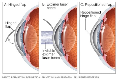
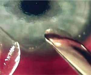
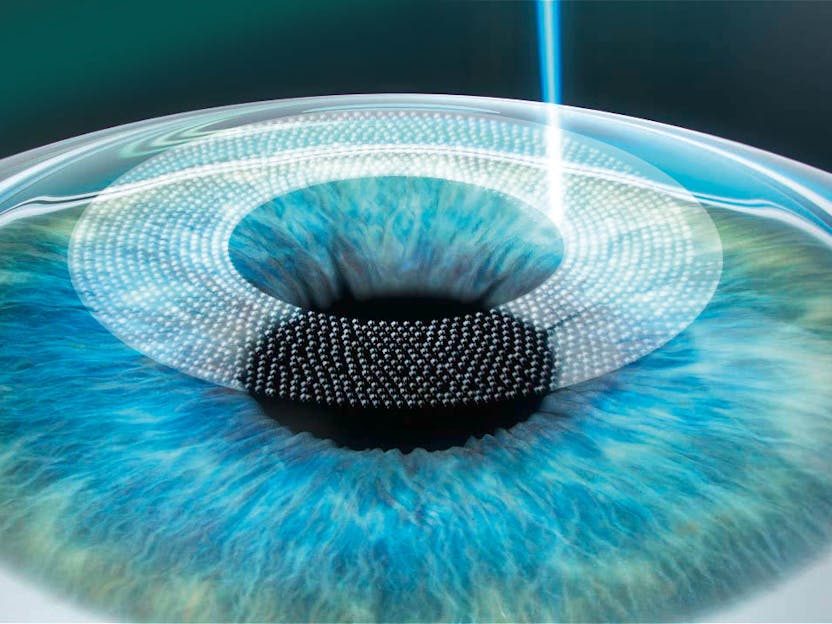
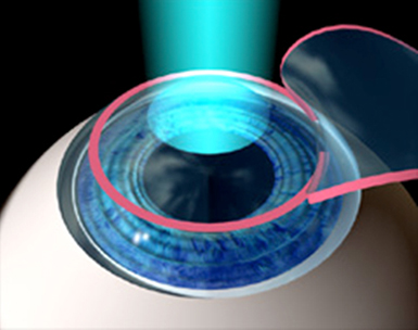
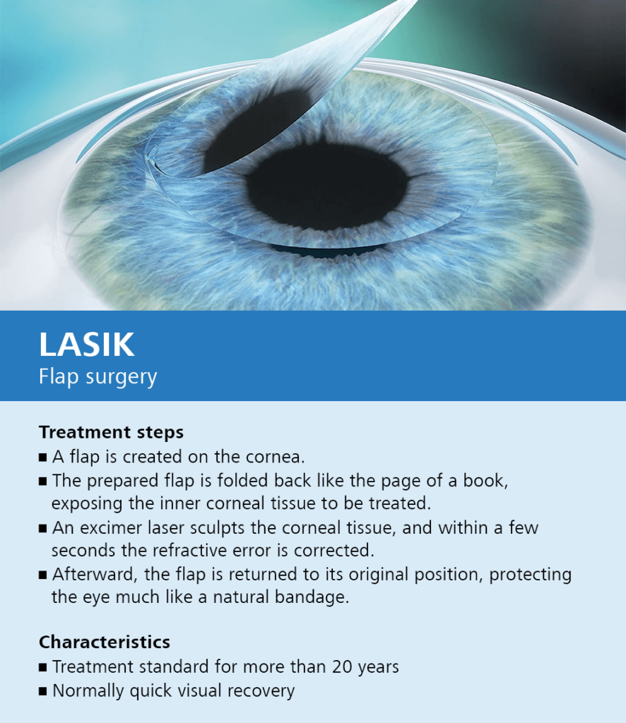
During LASIK eye surgery, the surgeon first administers numbing drops, which take effect in 1015 minutes, and then creates a thin, corneal flap with the laser The surgeon then pulls back the flap to expose the underlying tissue and reshapes the stroma layer of the cornea Then, the laser removes cells according to your prescription. The first step in the LASIK procedure is creating a corneal flap This is usually performed with a surgical tool called a microkeratome or a laser called IntraLase A ring is placed on the surface of the cornea and is held in place with suction. ILASIK combines the accuracy of the Excimer laser with the quick healing characteristics of a Femtosecond created flap Using a Femtosecond laser, a thin, protective flap of corneal tissue, attached by a hinge on one side, is folded back so the inner tissue of the cornea (stroma) can be treated with the excimer laser.
When performing all laser LASIK, our physicians use a gentle laser to create the corneal flap This technology, also used to facilitate laser cataract surgery, allows them to better customize the corneal flap for each patientAs the name states, no blades are used in this kind of laser eye surgery. The eye surgeon will then use a tiny blade to cut a hinged flap at the front of the cornea With the flap folded back, the surgeon will use a laser to remove precise amounts of tissue and remodel. How Does Lasik Work?.
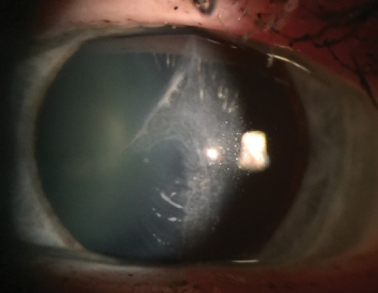
A hole appears in the middle of the flap Incomplete flaps. LASIK works by removing (ablating) tissue from the cornea This reshapes the surface of the cornea to correct the refractive error The amount of corneal tissue that is removed from the stroma is call the ablation depth, and this depends on the degree of refractive error. ILASIK combines the accuracy of the Excimer laser with the quick healing characteristics of a Femtosecond created flap Using a Femtosecond laser, a thin, protective flap of corneal tissue, attached by a hinge on one side, is folded back so the inner tissue of the cornea (stroma) can be treated with the excimer laser.
There are two steps of LASIK 1 Creation of a corneal flap to allow access to your underlying corneal tissue This is performed using the IntraLase femtosecond laser to create a precise, smooth flap in the safest manner possible 2 Corneal reshaping using Wavefront VISX CustomVue technology Only a miniscule amount of corneal tissue is gently. During LASIK eye surgery, the surgeon first administers numbing drops, which take effect in 1015 minutes, and then creates a thin, corneal flap with the laser The surgeon then pulls back the flap to expose the underlying tissue and reshapes the stroma layer of the cornea Then, the laser removes cells according to your prescription. The laser creates a corneal flap with edges that enables the flap to fit more securely in place after LASIK surgery, potentially reducing healing time An excimer laser is used to reshape the underlying corneal tissue to correct your vision, then the flap is returned to its original position The procedure typically takes about minutes.
Introduction Laser Assisted In Situ Keratomileusis (LASIK) flap complications occur more frequently in the early postoperative period 1 However, traumatic dislocations have occurred up to 14 years after the procedure 2 On the other hand, total avulsion of the LASIK corneal flap (sometimes with flap loss) is much less common This event has been related with intraoperative free caps but also. As a result, there is no way for the flap to remain Buttonhold flaps – occur when the flap is too thin;. February 4, 15 by Andrew Caster Myth A common concern about Lasik surgery is that the corneal flap will never heal To break it down quite simplygive it some time In the early days after Lasik, wounds should be handled with great care because that’s when the eye is most vulnerable and susceptible to trauma.
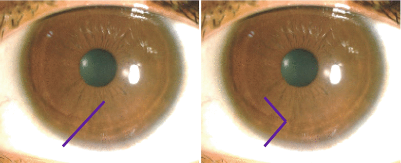
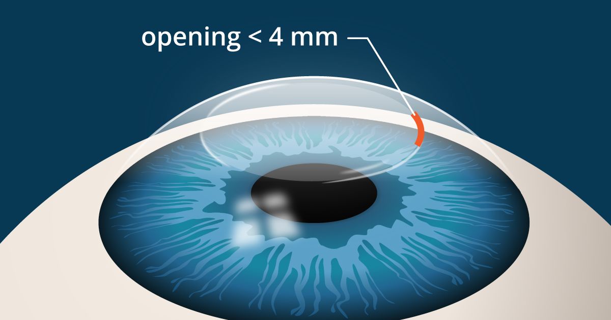
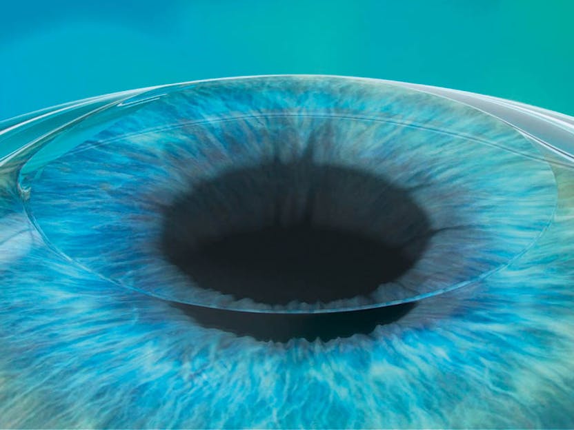
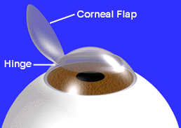
Corneal transplant LASIK permanently thins and thereby weakens the cornea This can lead to the need for a cornea transplant, despite a LASIK surgery being deemed “successful” Corneal flap dislocation The corneal flap is altered during every LASIK surgery However, in some cases, this eventually leads to a dislocated corneal flap. ILASIK combines the accuracy of the Excimer laser with the quick healing characteristics of a Femtosecond created flap Using a Femtosecond laser, a thin, protective flap of corneal tissue, attached by a hinge on one side, is folded back so the inner tissue of the cornea (stroma) can be treated with the excimer laser. Creating the Corneal Flap To create the corneal LASIK flap, the LASIK surgeon uses a handheld device or a laser to cut the top layers of corneal tissue at a predetermined depth One edge of the flap is left uncut, forming a hinge Using the hinge, the surgeon folds back the flap to access the underlying corneal tissue.
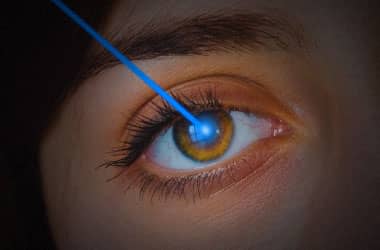
At the start of your LASIK surgery, an eye surgeon will use a femtosecond laser to create a flap on the surface layer of the cornea This laser is computerguided, allowing the surgeon to produce the flap with a perfectly precise circular incision around the outer corneal tissue that is catered to the specific eye shape. The creation of a flap in the outermost layer of the cornea is what distinguishes LASIK from other forms of laser refractive surgery, such as PRK and LASEK This means that flap complications are a potential risk of all forms of LASIK. Alright, so how does the corneal flap heal after LASIK?.
The first step to any laser vision correction procedure is to fold away the outer layer of the cornea, the part of the eye that both blocks foreign bodies and focuses light toward the retina Instead of completely removing a section of the cornea, LASIK technicians use a specially formulated tool to gently fold a sliver of the cornea back. Then the LASIK flap was repositioned and the cornea was continuously illuminated for 130 minutes with 30 mW/cm 2 using a KXL ultraviolet light source (Avedro Inc) delivering an energy dose of 2. Complications relating to the corneal flap created during LASIK include Free caps – occur when no hinge is made during flap creation;.
Intraoperative Complications Microkeratomerelated flap complications Other risk factors include loss of suction, defective blade, abnormal Corneal Epithelial Defect The risk factors of epithelial erosion during LASIK include older age, previous corneal Limbal Bleeding It occurs in two. The corneal flap heals in different ways at different parts of the flap We’ve broken it down for you here Epithelial layer Your epithelium is the very, very thin layer found on the surface of the cornea It begins healing almost immediately after the flap has been replaced and is the reason that you don’t feel a “line” between the flap and the rest of your eye. The corneal flap begins healing immediately after the LASIK procedure In fact, with the use of a LASIK flap, the corneal tissue can be as much as 90% healed within 24 hours During the first day or two after surgery, the outer surface of the cornea, known as the epithelium, seals the edges of the corneal flap Over the next few weeks, natural substances inside your cornea bond the corneal flap to the underlying tissue.
During LASIK surgery, a small flap is created in the frontal, topmost layer of the cornea This layer is called the epithelium Using this epithelial flap, the cornea can be reshaped and recontoured as needed to address refractive errors (ie, myopia/nearsightedness, hyperopia/farsightedness, and astigmatism). The LASIK flap heals back into its original position and the healing process is not limited to he edge alone The healed flap is stable and unlikely to dislodge with eye rubbing Significant eye trauma could potentially dislocate the flap, however, such instances are rare Flap creation making use of the femtosecond laser provides for even greater stability and reduced chances of flap movement or dislocation. Creation of thin, irregular corneal flap, and sometimes buttonhole flap which are the formation of holes in corneal flap Another complication that can occur is creation of small flap that gives insufficient size of corneal stromal bed for laser ablation These lasik complications can be treated by replacing the flap again and allow it to heal.
How Does Lasik Work?. Moreover, excessive flap manipulation may produce an irregular flap and cause corneal striae Mechanical trauma after LASIK can dislocate the flap and result in striae This trauma can be minimal, caused by eye rubbing or eye squeezing Although dry eye does not cause striae, an epithelial defect resulting from the condition can. PRK is not susceptible to eye injury As explained above, The LASIK flap and cornea never heal back to their original strength This flap, although sealed, can be dislodged or damaged by certain types of eye injuries This is the reason that the US military prefers surface ablation (PRK) to LASIK for most situations and military specialties.
There are two steps of LASIK 1 Creation of a corneal flap to allow access to your underlying corneal tissue This is performed using the IntraLase femtosecond laser to create a precise, smooth flap in the safest manner possible 2 Corneal reshaping using Wavefront VISX CustomVue technology Only a miniscule amount of corneal tissue is gently. The fact is that the corneal flap made in LASIK is selfhealing Initially, this vacuum effect keeps it in position, with the cells lining the inner surface of your cornea, known as endothelial cells, pumping water out to the inner part of the eye and the suction holding it in place During the first day or two after surgery, the outer surface. The corneal flap heals in different ways at different parts of the flap We’ve broken it down for you here Epithelial layer Your epithelium is the very, very thin layer found on the surface of the cornea It begins healing almost immediately after the flap has been replaced and is the.
The corneal flap begins healing immediately after the LASIK procedure In fact, with the use of a LASIK flap, the corneal tissue can be as much as 90% healed within 24 hours During the first day or two after surgery, the outer surface of the cornea, known as the epithelium, seals the edges of the corneal flap Over the next few weeks, natural substances inside your cornea bond the corneal flap to the underlying tissue. In LASIK for nearsightedness (myopic LASIK), the evaporation of the collagen lamellae is greater in the central part of the corneal bed and gradually lessens toward the flapbed in the periphery When the flap is laid down, the overall corneal curve is flatter. Laser Assisted In Situ Keratomileusis (LASIK) flap complications occur more frequently in the early postoperative period 1 However, traumatic dislocations have occurred up to 14 years after the procedure 2 On the other hand, total avulsion of the LASIK corneal flap (sometimes with flap loss) is much less common.
During LASIK surgery, a flap incision is created in the outer, epithelial, layer of the cornea With the flap open, the surgeon can then reshape the stromal layers of the cornea to achieve the precise shape upon which light can directly reflect off the retina and produce clear vision Why Is Corneal Thickness Important?. The emergence of femtosecond laser technology has revolutionized lamellar flap creation The refractive surgery community continues to debate whether to use either a blade or laser to create corneal flaps This article discusses the clinical advantages and disadvantages of the femtosecond laser and mechanical microkeratome for LASIK flap creation. How the flap is made during laser eye surgery When laser eye surgery was approved by the FDA for use in 19, the surgeon would use a microkeratome – a surgical instrument with an oscillating blade – to precisely create the corneal flap In 01, the femtosecond laser was approved for use in laser eye surgery This laser performs a perfect incision by using ultrashort pulses.
/Femto%20LASIK%20Step%201.png)
Refractive Surgery Lasik

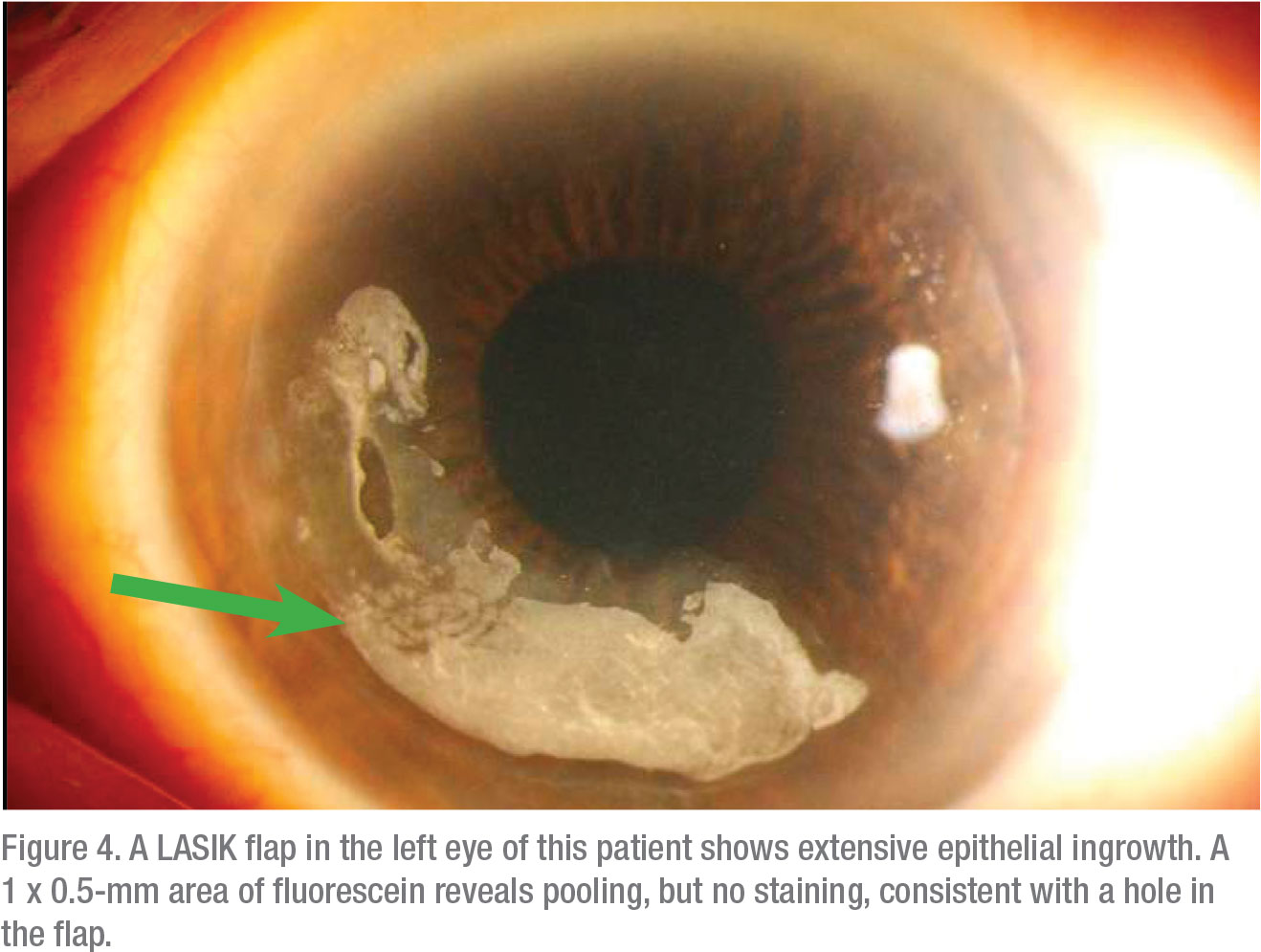
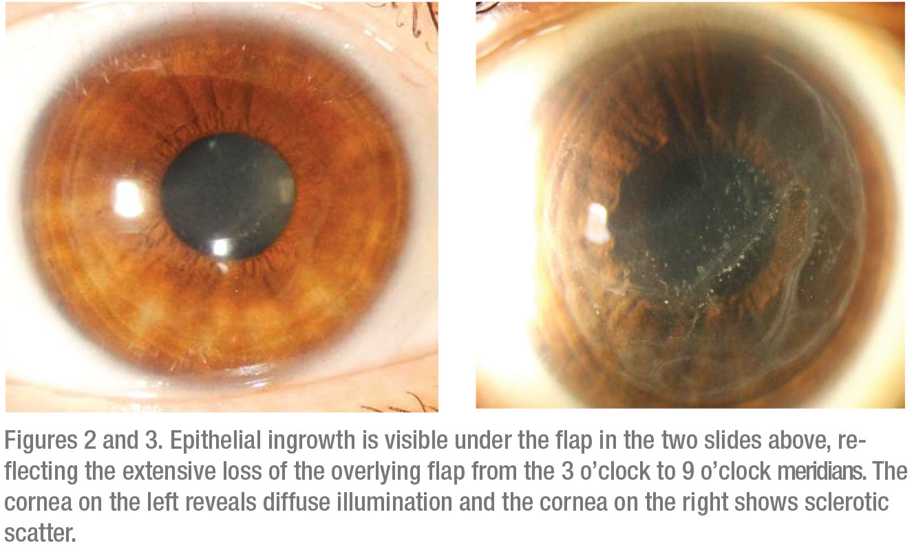
Epithelial Ingrowth Following Laser In Situ Keratomileusis Lasik Prevalence Risk Factors Management And Visual Outcomes Bmj Open Ophthalmology

Eyeworld Bad Memories Of Flap Complications
.jpg)
Lasik Flap Dislocation The Flap Never Heals
Lasik Eye Surgery Mayo Clinic

Late Lasik Flap Study Eurotimes

Eyeworld Bad Memories Of Flap Complications
Laser In Situ Keratomileusis Lasik American Academy Of Ophthalmology

Lasik Flap Versus Surface Prk No Flap Western Laser Eye Associates

Lasik Eye Surgery Creationwiki The Encyclopedia Of Creation Science
Lasik

Lasik Laser Services Gupta Eye Hospital
Lasik Wikipedia

Eye Health Pain Free Lasik Surgery Is Often Best Most Common Solution For Poor Vision Kyforward Com Kyforward Com
Corneal Compromise How To Assess The Risk Of Post Lasik Ectasia

Pdf One Case Which Injury Lead To Corneal Flap Laceration After Lasik Nine Years Ago Semantic Scholar

Refractive Surgery Lasik Department Of Ophthalmology Nus Yong Loo Lin School Of Medicine
/detached-lasik-flap(640).jpg)
Lasik Flap Dislocation The Flap Never Heals

Hd Corneal Flap Displacement From Eye Trauma After Lasik Em Rap
Smile Eye Surgery What S Different Than Lasik

Management Of A Traumatic Flap Dislocation Seven Years After Lasik

Late Post Traumatic Flap Dislocation And Macrostriae After Laser In Situ Keratomileusis Sinha R Shekhar H Tinwala S Gangar A Titiyal Js Oman J Ophthalmol
How To Handle Lasik Rip Offs
Laser In Situ Keratomileusis Lasik American Academy Of Ophthalmology
Lasik Laser Eye Surgery

Lasik Eye Surgery

Full Text Traumatic Corneal Flap Displacement After Laser In Situ Keratomileusis Imcrj

Escrs Journal Of Cataract And Refractive Surgery

Troubleshooting In Lasik Eye News
Before Enhancing Post Lasik Patients

Before Enhancing Post Lasik Patients

Lasik Flap Surl0001 Stock Eye Images

Eyeworld Management Of Rare Post Lasik Complications
Laser Refractive Surgery Lasik Lasek Prk And Ptk Discovery Eye Foundation
Lasik Center In Boston Ma Laser Vision Correction Boston Laser
My Eye 6 Days After Lasik Eye Surgery Corneal Flap Healing Red Spots Fading Youtube
Does Corneal Flap Thickness Matter In Lasik

How Does Cornea Thickness Affect My Candidacy For Lasik Price Vision Group

Lasik Flap Made With Femtosecond Laser Youtube

Full Text Traumatic Corneal Flap Displacement After Laser In Situ Keratomileusis Imcrj

The Risk Of Flap Complications After Lasik Eye Surgery Maida Customvision
Crstoday Vision Loss After Post Lasik Trauma

Late Traumatic Lasik Flap Loss During Contact Sport Sciencedirect
.jpg)
Lasik Flap Dislocation The Flap Never Heals
Expert Opinion Laser Eye Surgery Gponline
Lasik Laser Eye Surgery
Before Enhancing Post Lasik Patients

Lasik Eye Surgery Mayo Clinic
Lasik Guider Laser Vision Correction Guide Does The Lasik Flap Ever Heal Completely
Understanding The Risk Of Lasik And Flap Complications

Lasik Complications And Their Management Ento Key

Characterisation Of Corneal Fibrotic Wound Repair At The Lasik Flap Margin British Journal Of Ophthalmology
Lasik Know The Rewards And The Risks
The Long Term Effects Of Lasik Good And Bad Nvision Eye Centers
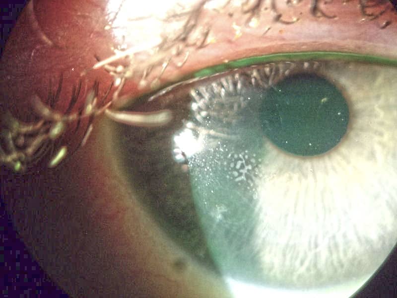
Epithelial Ingrowth Under Lasik Flap
Millennialeye Post Lasik Fungal Flap Infection

Lasik Interface Primary Complications

Lasik Eye Surgery In Santacruz Mumbai Samarth Eye Care

Lasik Photo Gallery Sclerallens Com

Early Post Lasik Flap Amputation In The Treatment Of Aggressive Branching Keratitis A Case Report
Q Tbn And9gcsuqogvtezopkkbaabxhlouhhh4fbcqyb9wrltk7rogroagnb8g Usqp Cau
Lasik Complications Epithelial Ingrowth Risk At 50 Endmyopia
.jpg)
Epithelial Ingrowth Lasikcomplications Com
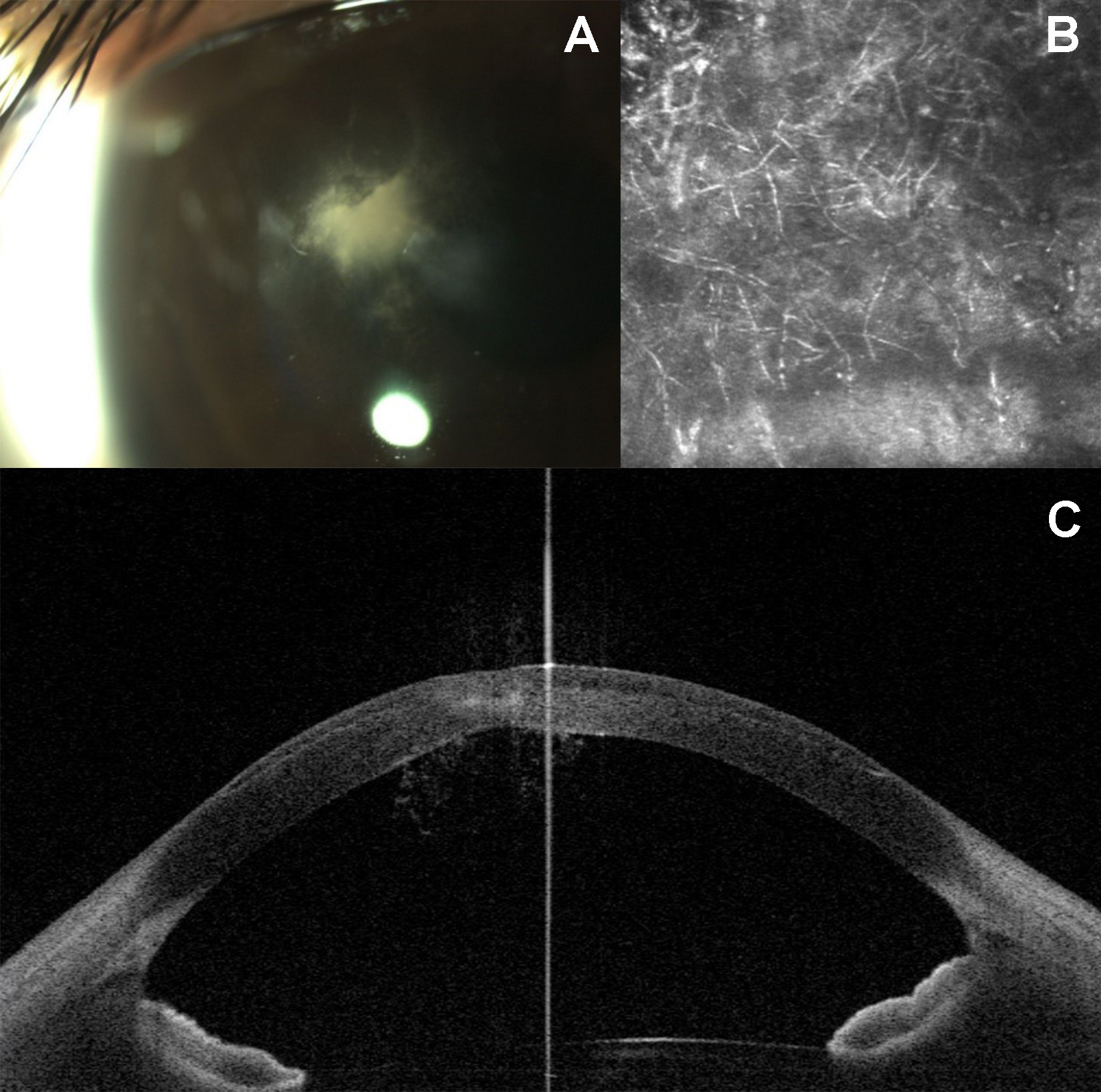
/ectasia_flap_margin(425).jpg)
Lasik Flap Dislocation The Flap Never Heals

Flap Striae Columbia Ophthalmology

Flap Striae After Lasik Can Be Treated Successfully
Q Tbn And9gctxxqxjjpvvi4xlgrxpldxjizal5f2ir T5evlptk1oe3wovlvu Usqp Cau

Long Term Outcomes Of Flap Amputation After Lasik

Traumatic Flap Dislocation 10 Years After Lasik Case Report And Literature Review Sciencedirect

The Lasik Evolution
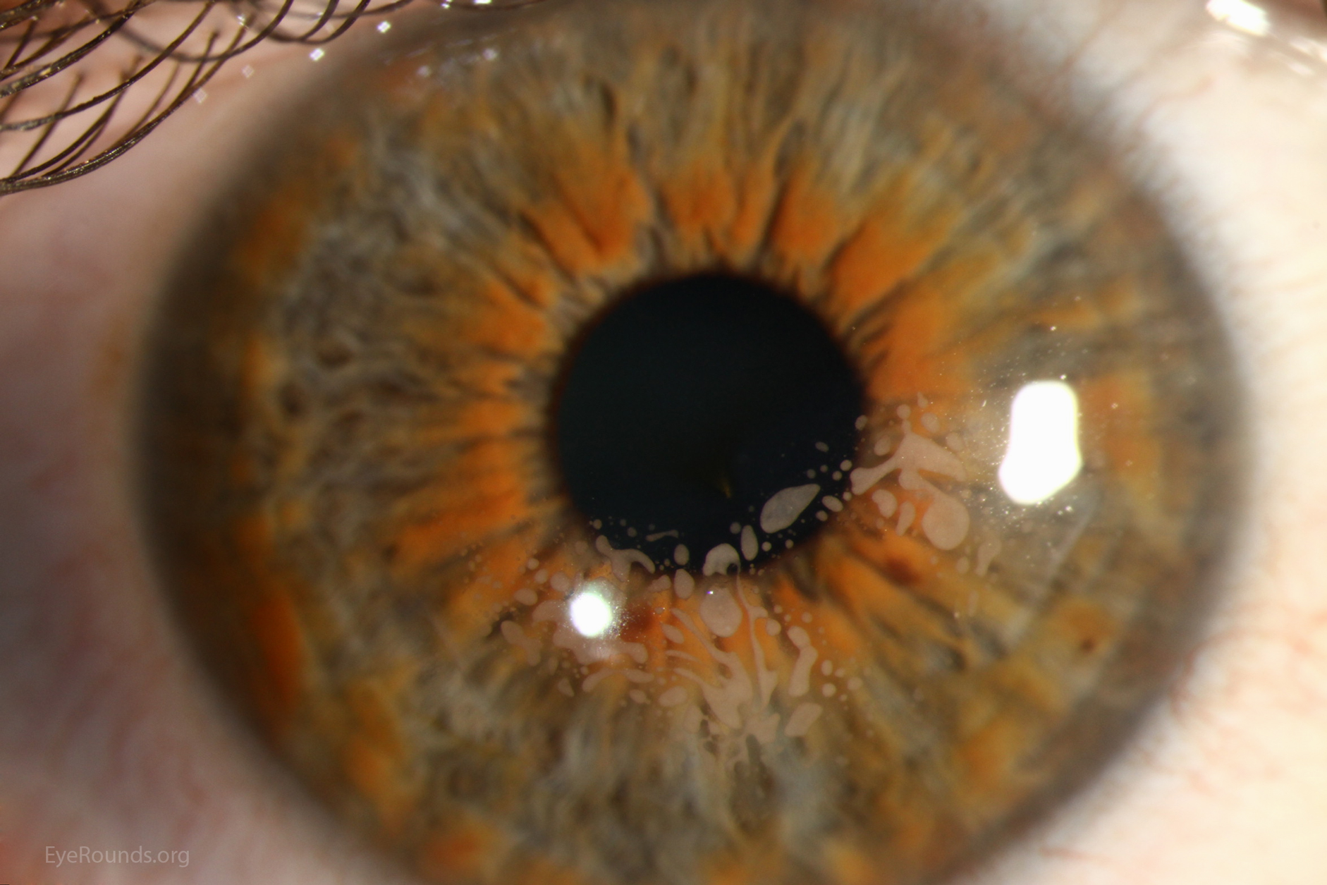
Epithelial Ingrowth Under Lasik Flap

Eyeworld Management Of Rare Post Lasik Complications
Lasik Eye Surgery Series Procedure Part 2 Medlineplus Medical Encyclopedia

Post Lasik Granular Corneal Dystrophy

What Can Go Wrong Lasik Surgery Wright Vision Center Wright Vision Center
/PRK%20Step%202.png)
Refractive Surgery Lasik

Eye에 있는 핀
Lasik Prk Grosinger Spigelman Grey
Lasik Clearvision Eye Clinic Lasik Centre
Lasik Flap Everything You Need To Know Iq Laser Vision

The Para Position Of The Corneal Flap In The Left Eye Is Satisfactory 2 Download Scientific Diagram

Replacing My Corneal Flap During Lasik Surgery Interestingasfuck

Case Study Enhancement 13 Years After Lasik Consult Qd
Q Tbn And9gcs7ligxsexqrccrqe0agwsttwsj67egyvhguue35vutunvq1wav Usqp Cau

Lasik Enhancement With Epithelial Cells Growing Under Flap Youtube
Lasik Surgery Explained Definition Procedure Results
Kadam Eye Lasik Centre Lasik Surgery With Flap Creation

Pdf Management Of Slipped Laser In Situ Keratomileusis Flap Following Intrastromal Corneal Ring Implantation In Post Lasik Ectasia

Lasik Prk
Prk Vs Lasik Eye Care Associates

Clinical Case With Post Lasik Ectasia Due To A Thick Corneal Flap A Download Scientific Diagram

Laser Refractive Surgery Lasik
Lasik Beverly Hills Lasik Eye Surgery Beverly Hills Gaster Eye

Lasik Surgery Explained Definition Procedure Results
Prk Vs Lasik Differences Pros Cons And What To Expect

Lasik Prk Grosinger Spigelman Grey
Q Tbn And9gcq 3vda4gl9aihbd2 Slfkmvh8yfq4ul Loc9hsvxwnwo0v Ld2 Usqp Cau
My Lasik Eye Surgery Experience This Article Is Not Written By A By Kavishka Gamage Medium


