Corneal Flap Dog

Conjunctival Pedicle Grafting Of The Cornea Information

Conjunctival Flap Cover Surgery 10 Year Review Yao Annals Of Eye Science

Case Of The Week
Surgery Of The Cornea And Sclera Veterian Key
How To Do Eye Surgery In Deep Corneal Ulcerated Dogs Youtube

Corneal Ulcer Treatment Dog Eye Ulcer Treatment Seah
The most common cause of corneal ulcer in dogs is by blunt trauma This includes your dog rubbing their eye on the carpet, getting scratched by a cat or any other contact where the cornea was hurt by a hard object It could've even been by a chemical burn in the cornea, due to a shampoo, drywall dust, etc.
Corneal flap dog. Young dogs can also develop ectopic cilia, or abnormal hairs that protrude from the conjunctiva lining the eyelid When the animal blinks, these cilia contact the cornea and create ulceration Surgery, typically under an operating microscope, is required to remove the ectopic cilia and allow the ulcer to heal. Enucleation should be considered in cases such as these with a grave prognosis for vision and comfort. Corneal ulcer is a condition that may affect dogs of all ages Chronic ulcers may be more common in middle aged and senior dogs The ulcers may be caused by injuries, bacterial, viral or fungal infections Treatment should be applied, as these ulcers may affect the vision of the pet and may also cause blindness.
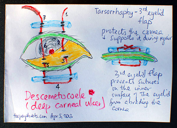
In one study, 48 corneal samples from dogs with SCCEDs taken during therapeutic superficial keratectomies demonstrated that the corneal tissue adjacent to the ulcerated area had epithelium that was not only poorly attached to the underlying stroma but also had abnormal epithelial structure11 The adhesion complexes between the epithelium and its basement membrane were often deficient or absent in the areas of and surrounding the ulceration11 Particularly interesting was the presence of an. My dog's conjunctival flap surgery?. In this VETgirl online veterinary continuing education video, we discuss the use and placement of a 3rd eyelid flap in your veterinary patient A third eyelid flap is an easy and quick procedure, and is a very good option for certain cases The 3rd eyelid can serve as tectonic support and protection for severe, melting corneal ulcers or large corneal perforations when a conjunctival graft is not possible.
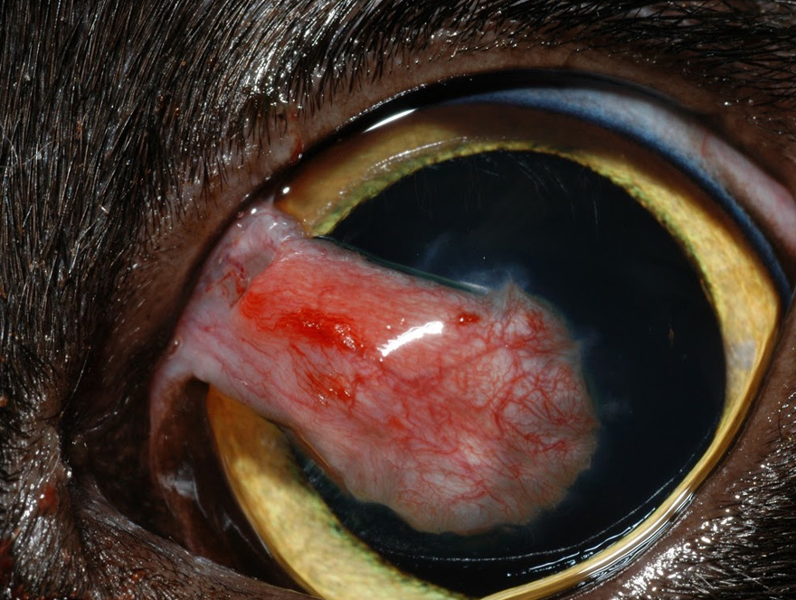
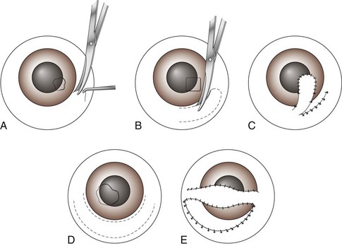
Dr David Williams discusses corneal ulcers and reports on a new treatment for an ulcer extending into the stroma and without a predisposing cause in dogs which are not candidates for surgery IT was the end of a great meeting in Denmark I had been lecturing to Danish veterinary ophthalmologists and enjoying their great hospitality too. Dissected flaps of conjunctiva offer a valuable means of covering the ulcerated cornea either temporarily or permanently I shall enumerate the corneal conditions for which they are most useful and shall describe the technique that I have used in applying them to the cornea Conjunctival flaps. The most common type of corneal restructuring graft is the conjunctival pedicle/advancement flap The techniques are suited primarily for restoring the corneal strength (marginal at best though), but is a very poor optical choice These grafts rarely result in clarity (Fig 2).
Examples of other diseases include epithelial dystrophy a weakening of the cornea which can be inherited in breeds such as Boxer Dogs drying of the cornea due to decreased tear production, called keratoconjunctivitis sicca (KCS or "dye eye") endocrine diseases such as diabetes mellitus, Cushing's. A corneal ulcer is the deep erosion of the eye’s third layer, which results in a cloudy appearance and pain for your pet It is commonly caused by trauma – either via the eyelashes rubbing against the eye (entropion), via a cat scratch or via contact with a sharp object. To report and compare the success rate of a conjunctival pedicle flap (CPF) alone vs a CPF with an underlying acellular submucosa implant for the repair of deep or perforating corneal wounds in dogs.
Purpose To evaluate the efficacy of superficial keratectomy and conjunctival advancement hood flap (SKCAHF) for the treatment of bullous keratopathy in canine patients Methods Nine dogs (12 eyes) diagnosed with progressive corneal edema underwent superficial keratectomy followed by placement of conjunctival advancement hood flaps. Corneal ulcers are one of the most common eye problems that we see in small animal medicine Common signs of corneal ulcers in dogs and cats include squinting, redness in the sclera (the part of the eye that is normally white), abnormal discharge from the eye – either watery or mucousy – and sometimes cloudiness or haziness to the eye itself. A corneal ulcer is a wound or break in the outermost layer of the cornea It is painful to the dog Causes of Canine Corneal Ulcers Eye ulcers in dogs may be caused by a wound to the eye This could be a scratch, abrasion, puncture, or other trauma Ulcers may be selfinflicted if the dog is rubbing his eye for any reason, including allergies.
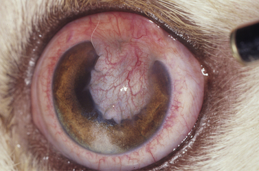
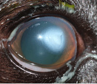
Specific causes of focal edema include focal corneal epithelial dysfunction (due to corneal ulceration), or focal endothelial cell damage (due to blunt corneal trauma, lens luxation, or keratic precipitates) (Figure 1) 2 FIGURE 1 Focal corneal edema secondary to corneal ulceration in a dog The pupil has been pharmacologically dilated. Thermal keratoplasty (a technique for reshaping the cornea) and Gunderson flap (replacing the damaged section of cornea with a section or “flap” of the patient’s own conjunctiva) procedures for corneal edema (swelling) associated with corneal endothelial degeneration (a condition in dogs that affects corneal clarity). After completing the conjunctival flap dissection, create the partialthickness corneal–scleral trabeculectomy flap, which is the guarded part of this procedure If a limbalbased technique was used, gently reflect the conjunctival flap over the cornea and gently hold it with smooth forceps or a WeckCel sponge.
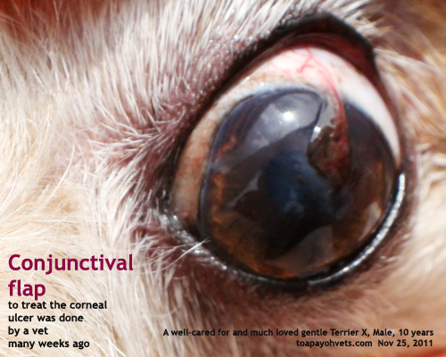
Corneal ulcers are a common problem in pets, and we offer a variety of treatments, including conjunctive flap grafting surgery The Conjunctival Flap or Graft Procedure Conjunctival flap grafting is one option for the treatment of deep corneal ulcers The conjunctiva is the pale pink tissue that covers the “white” of your pet’s eye. Symptoms of Corneal Dystrophy in Dogs White or grayish cloud in the center of the eye Irritated eyes. Corneal laceration with retinal detachment, dog Large, ventral corneal laceration with intraocular hemorrhage and retinal detachment (by Bscan ultrasonography) would not result in restoration of vision even if repaired;.
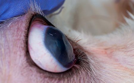
Corneal Endothelial Dystrophy The hallmark sign of corneal endothelial dystrophy in dogs is eyes with a blue or foggy appearance As the disease progresses, patients experience ocular discomfort and pain Your pet may also avoid bright light or show signs of visual discomfort when outdoors. The size of the nearly round to oval cornea (vertical/horizontal) varies by animal species dog (85 × 95 mm), cat (84 × mm), horse (166 × 179 mm), and cow (152 × 164 mm) The animal cornea consists of the superficial epithelium and basement membrane, large and relatively acellular stroma, deeper Descemet membrane, and deep single layer endothelium. Corneal dystrophies (abnormal form or structures) that occur in both eyes are likely inherited or breedpredisposed in dogs, and often consist of triglyceride and cholesterol deposits within the connective tissue of the cornea Treatment for these deposits is not usually necessary unless they affect vision or become irritating.
Corneal ulcers are eye problems resulting from a loss in the tissue layers of your pet’s cornea, or lens This eye problem results in a pain and can impact their vision There are many potential causes for corneal ulcers such as trauma, disease, foreign body, or infection. Diffuse corneal edema in a dog with a penetrating corneal cat claw injury and secondary uveitis The wound from the cat claw can be seen on the ventromedial paraxial cornea Treatment involves emergency surgical support—either conjunctival grafting or placement of a third eyelid flap 4,5 FIGURE 3 Feline acute bullous keratopathy Note. The most severe complications postoperatively was conjunctival flaps dissolving and corneal perforation for 3 eyes out of 253 patients (12%), and corneal transplantation had to be performed to avoid enucleation In addition, 4 eyes out of 253 patients (16%) presented cystic flap and two of them received surgical excision.
The latter is “almost like a rubbery piece of tissue that, in some ways, is even a little easier to dissect than a partialthickness corneal piece,” said Dr Karp Irradiated sterile cornea also has a long shelf life and is applicable to nonendothelial procedures 2 It’s also possible to temporarily transplant other types of banked tissue. Corneoconjunctival transposition flap is a form of autogenous corneal transplant in which clear corneal tissue attached to adjacent bulbar conjunctiva is elevated and slid into a central corneal defect, resulting in a clearer visual axis and better visual outcome It also has the advantage of physically filling the defect and providing rapid wound strength. Corneal ulcers are one of the most common eye problems that we see in small animal medicine Common signs of corneal ulcers in dogs and cats include squinting, redness in the sclera (the part of the eye that is normally white), abnormal discharge from the eye – either watery or mucousy – and sometimes cloudiness or haziness to the eye itself.
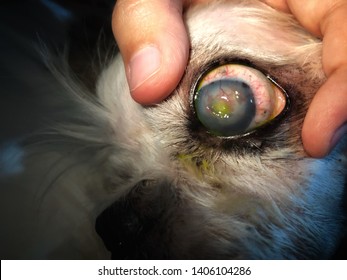
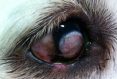
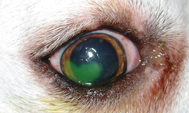
Nonhealing Superficial Corneal Ulcers in Dogs by Daniel Biros, DVM, DACVO Sometimes loose flaps of epithelial sheets or fragments of epithelia from the wound edges are seen freely hanging from the ulcer’s edge, misguided and unsuccessful attempts at wound healing Most cases are unilateral, but bilateral ulcers may present. Conjunctival flap cut scenes 犬のデスメ膜瘤保護のための結膜フラップ作成シーン part 1 は結膜弁の切り出しシーン。. Treating corneal ulceration in dogs part 2 deep ulcers Author Mateusz Jaksz, Claudia Busse Categories Companion animal, Vets Date March , 17 Deep corneal ulcers are defined by a defect of the epithelium and stromal loss that may extend as far as the Descemet’s membrane Figure 1a Deep corneal ulcer in two pugs, frontal view.
These erosions usually occur in middleaged dogs (ie, 7 to 9 years) and in all breeds of dogs, although Boxers are often overrepresented Diagnosis A spontaneous, chronic, corneal epithelial defect should be suspected in any middleaged dog with a nonhealing corneal erosion (ie, an uncomplicated erosion that has not healed within 1 to 2 weeks). Deep corneal ulceration and corneal grafting procedures Your dog has been diagnosed with a deep corneal ulcer and a grafting procedure has been recommended The information below will help you to understand your options and what to expect following the operation. Large Replacement Dog Door Flap Compatible with PetSafe Freedom Doggie Doors PAC Measures 10 1/8" x 16 7/8" Made from flexible, durable, weather resistant materials Doggie Door Flap 45 out of 5 stars 1,928 $2699 $ 26 99 ($2699/Count) Get it as soon as Fri, Jan 22.
The cornea is the transparent window of the eye that allows light to enter Damage to the surface of the cornea (most commonly by trauma) exposes the underlying tissue, causing an ulcer If the ulcer is shallow and uncomplicated it will usually heal within a few days without surgery. Superficial keratectomy and 360° conjunctival flap for bullous keratopathy in a dog A case reportpdf (Milvago chimachima) treated with a modified third eyelid flap 1 tion of the cornea. Endothelial corneal dystrophy 1Swelling of the cornea with fluid blisters 2Possible impairment of the vision 3Middleaged dogs are at higher risks 4Also affects young animal Dogs that are at higher chances of developing corneal dystrophy 1Boston terriers 2Chihuahuas 3Dachshunds 4May affect other breeds Diagnosis and Tests.
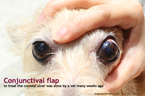
As well, corneal abrasions can lead to other issues, including corneal ulcers or keratitis The vet will administer a special dye to the dog's eye and look for damaged eye tissue If a foreign body is in his eye, the vet will remove it and may administer medication, such as an antibiotic ointment or drops to prevent infection. The aim of this study was to report a case of BK in a dog and the complete recovery of the ocular structure and visual function, with a third eyelid flap associated with the use of autologous blood serum topicallyCase A 2yearold Shih Tzu male dog, weighing 43 kg, with recurrent bilateral eye discomfort was brought to Ophthalmologist Veterinarian Assistance. This consists of making a flap of tissue from the conjunctiva (pink tissue surrounding the eye) and suturing it over the affected areas of the cornea This procedure relieves the discomfort caused by corneal ulceration, however it will leave a scar on the cornea, and does not address the main cause of the corneal changes.
In one study, 48 corneal samples from dogs with SCCEDs taken during therapeutic superficial keratectomies demonstrated that the corneal tissue adjacent to the ulcerated area had epithelium that was not only poorly attached to the underlying stroma but also had abnormal epithelial structure11 The adhesion complexes between the epithelium and its basement membrane were often deficient or absent in the areas of and surrounding the ulceration11 Particularly interesting was the presence of an. Pigmentation and scarring of the cornea are common postoperatively, particularly in dogs of brachycephalic (shortnosed) breeds To reduce scarring, we often dispense a course of eye ointment (‘Optimmune’) following the surgery. She had it done because of a corneal ulcer and the vet wanted a blood supply to get in there i'm just not sure what to expect after the surgery i've called the vet with what i thought were changes but they said i only need to worry if she's squinting or keeping her eye.
Pet Door Frame Durable Easy Install Dog Doggie Flap Mount Secure Medium White 49 out of 5 stars (28) Total Ratings 28, 100% agree Would recommend $5563 New PetSafe PAC XL Premiun Replacement Flap 48 out of 5 stars (23) Total Ratings 23, 100% agree Would recommend $5495 New. Corneal Endothelial Degeneration (CED) is a degenerative condition in dogs that affects the clarity of the cornea This agerelated disease can result in blindness and severe ocular pain from secondary complications The cornea is the clear window at the front of the eye It is a very delicate structure which is less than a millimeter thick. Now the symptoms can vary depending on the type of dystrophy we’re talking about but some of the symptoms might be Discoloration of eyes or rings in the cornea Corneal Lipidosis If your dog has yellowish eyes then it could be corneal lipidosis This can be caused by corneal If your dog has.
In the present case, lamellar keratectomy associated with third eyelid flap was effective in the treatment of corneal epithelial inclusion cyst in a dog Appearance of corneal cyst in the left. Apply local anaesthesia, and disinfect with liberal use of 1% topical povidone Use a sterile cotton swab to debride all the loose epithelium In some cases you will strip all of the corneal epithelium If the dog is cooperative a grid keratotomy can be done with a nurse holding the dog. Nonhealing Superficial Corneal Ulcers in Dogs by Daniel Biros, DVM, DACVO dbiros@angellorg angellorg/eyes Otherwise known as spontaneous chronic corneal epithelial defects (SCCEDs), these clinical cases often frustrate the clinician because the normal woundhealing process for superficial corneal ulcers is thwarted, and often the affected dogs have prolonged periods of discomfort and in some cases progressive keratitis despite aggressive medical treatment.
The latter is “almost like a rubbery piece of tissue that, in some ways, is even a little easier to dissect than a partialthickness corneal piece,” said Dr Karp Irradiated sterile cornea also has a long shelf life and is applicable to nonendothelial procedures 2 It’s also possible to temporarily transplant other types of banked tissue. Daniel M Dorbandt, Phillip A Moore, Kathern E Myrna, Outcome of conjunctival flap repair for corneal defects with and without an acellular submucosa implant in 73 canine eyes, Veterinary Ophthalmology, /vop, 18, 2, (), (14). There are several causes of corneal ulcers in dogs "The most common cause is trauma" The most common cause is trauma An ulcer may result from blunt trauma, such as a dog rubbing its eye on the carpet, or due to a laceration, such as a cat scratch or a contact with a sharp object.
Has anyone had a pet go through this?. Corneal ulceration is one of the most common eye diseases in domestic dogs, and is a major cause of blindness due to either scarring or corneal perforation Damage to the cornea can cause substantial pain Corneal ulcers vary in severity, and can be classified into grades based on their depth. Nonhealing Superficial Corneal Ulcers in Dogs by Daniel Biros, DVM, DACVO Sometimes loose flaps of epithelial sheets or fragments of epithelia from the wound edges are seen freely hanging from the ulcer’s edge, misguided and unsuccessful attempts at wound healing Most cases are unilateral, but bilateral ulcers may present.
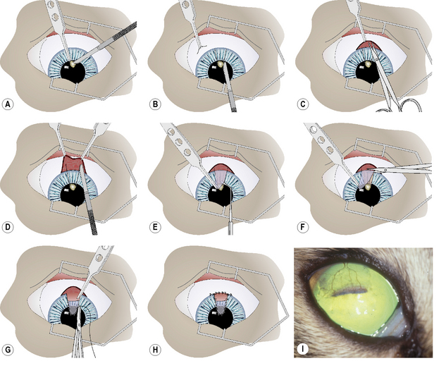
Diffuse corneal edema in a dog with a penetrating corneal cat claw injury and secondary uveitis The wound from the cat claw can be seen on the ventromedial paraxial cornea Treatment involves emergency surgical support—either conjunctival grafting or placement of a third eyelid flap 4,5 FIGURE 3 Feline acute bullous keratopathy Note. THE TECHNIQUE of using a conjunctival flap for the treatment of chronic corneal ulceration was described by Gunderson 1 in the late 1950s and became a standard surgical procedure There are some drawbacks to this procedure, however 1,2 During the past years, progress in microsurgical technique, aided by the microscope and fine surgical tools, has enabled us to use a selective pedunculated.

Third Eyelid Flap In Dog Youtube

Conjunctival Pedicle Flap Following Removal Of Corneal Sequestrum From A Cat S Eye Corneal Eyes Cats
Surgery Of The Cornea And Sclera Veterian Key

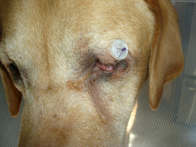
My Dog Had A Third Eye Lid Flap Today Around 3 00pm I Picked Her Up St 5 00 She Has The Cone Collar That Is Really

Pin By Cullenwebb Animal Eye Speciali On Eye Diseases Corneal Ulcer Corneal Eyes

Corneal Ulcers In Animals Wikipedia
Diagram Of Transpalpebral Third Eyelid Flap Note How The Suture Is Download Scientific Diagram

Use Of Porcine Small Intestinal Submucosa For Corneal Reconstruction In Dogs And Cats 106 Cases Goulle 12 Journal Of Small Animal Practice Wiley Online Library
Canine Dog Veterinary Surgery Anaesthesiaveterinary Surgery Anaesthesia Singapore Toa Payoh Vets Hamster Medicine Surgery Cases Health Sickness Singapore Singapore Toa Payoh Vets
Corneal Disease Inherited In Dogs Petmd
Veterinary Conjunctival Flap Graft Animal Eye Consultants

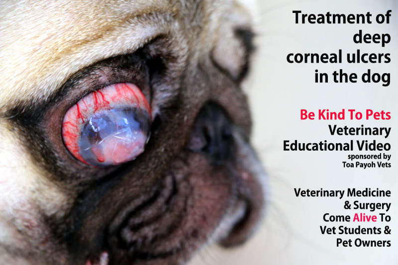
Deep Corneal Ulceration And Corneal Grafting Procedures
Www Agriculturejournals Cz Publicfiles 139 17 Vetmed Pdf
4kzix Cuadobbm

A Modified Technique Of Keratoleptynsis Letter Box For Treatment Of Canine Corneal Edema Associated With Endothelial Dysfunction Giannikaki Veterinary Ophthalmology Wiley Online Library

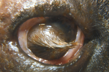
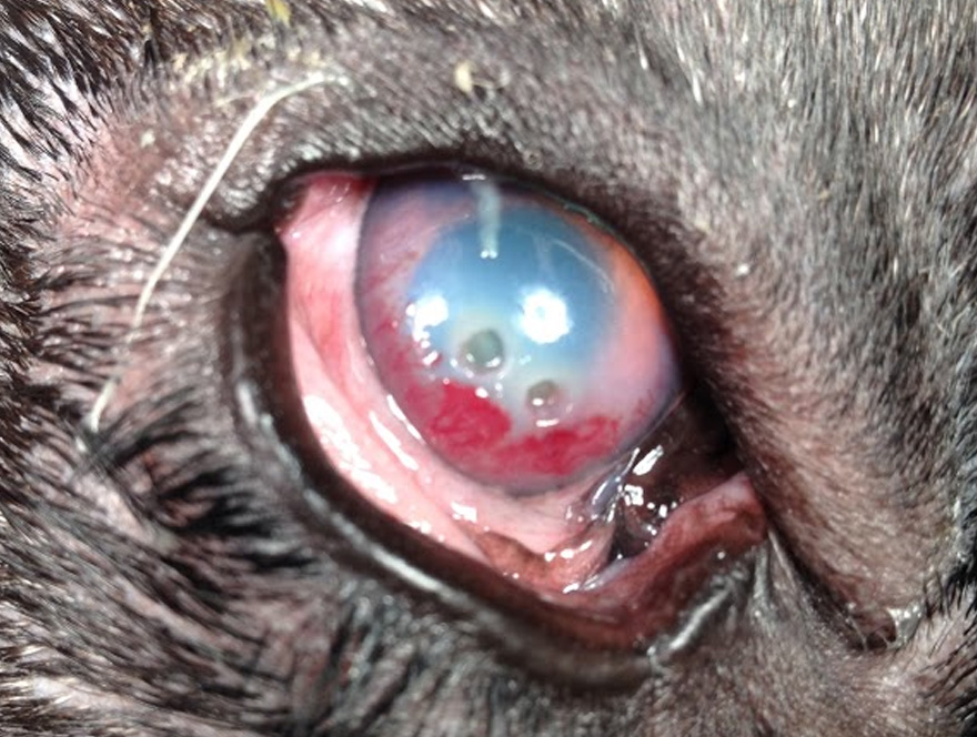
Deep Stromal Corneal Ulcers Descemetocele And Iris Prolapse In Animals Emergency Medicine And Critical Care Veterinary Manual

Cornea Eye Diseases And Disorders Merck Veterinary Manual

Corneal Epithelial Inclusion Cyst In A Dog
The Dog House Cornea Repair With Third Eyelid Flap Again

Deep Stromal Corneal Ulcers Descemetocele And Iris Prolapse In Animals Emergency Medicine And Critical Care Veterinary Manual

Clinical Case Challenge Blepharospasm Of The Right Eye News Center At Cummings School Of Veterinary Medicine At Tufts University
Eye Scratch Injuries In Puppiesthe Veterinary Expert Pet Health

Lasik Know The Rewards And The Risks
Q Tbn And9gcsi2blomwvzg H3kvhtrfvbq1fblbxnqf655l117qbzejibszli Usqp Cau
Www Vettimes Co Uk App Uploads Wp Post To Pdf Enhanced Cache 1 Treating Corneal Ulceration In Dogs Part 2 Deep Ulcers Pdf
Www Vettimes Co Uk App Uploads Wp Post To Pdf Enhanced Cache 1 Treating Corneal Ulceration In Dogs Part 2 Deep Ulcers Pdf

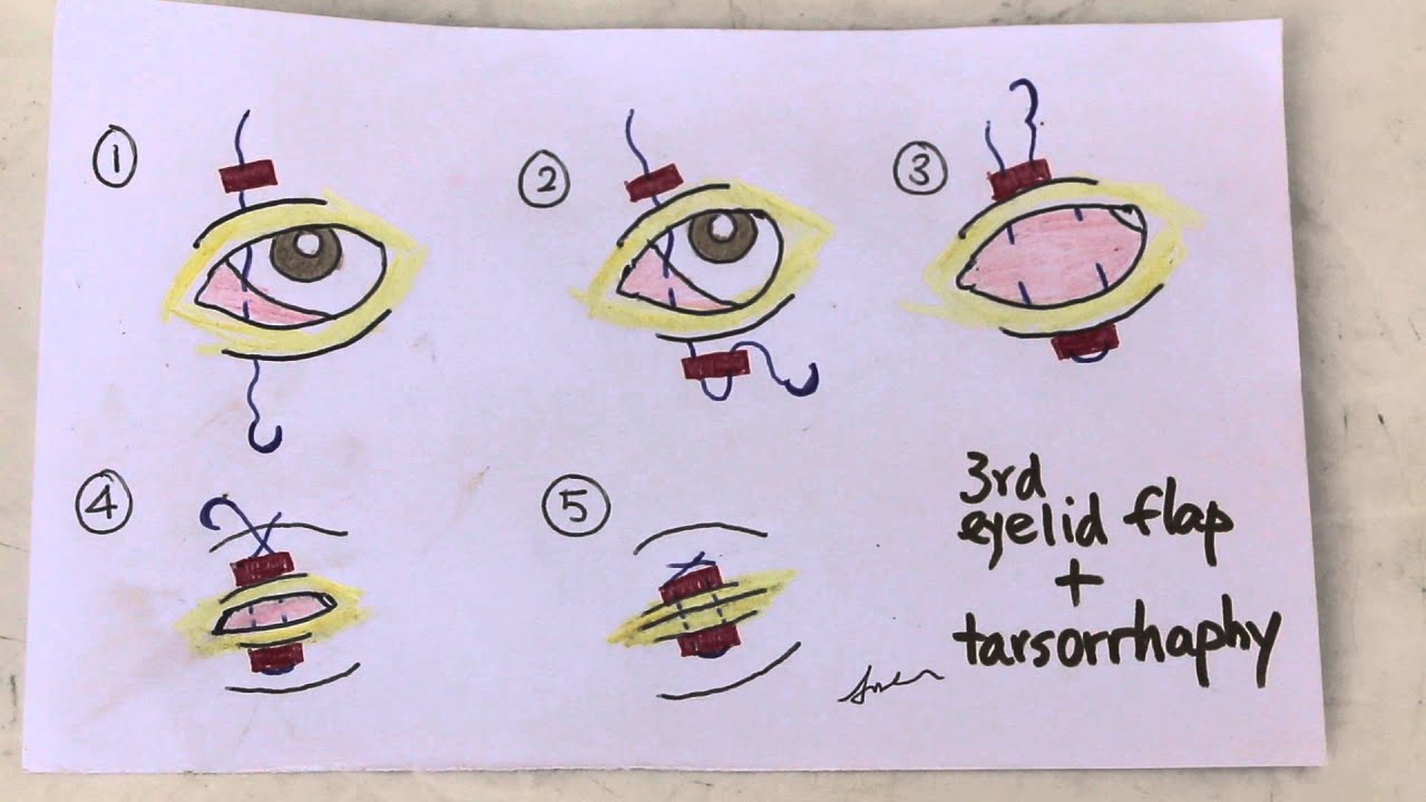
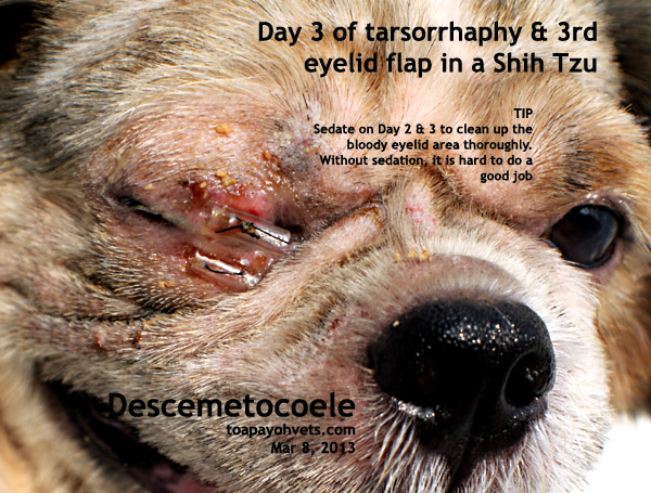
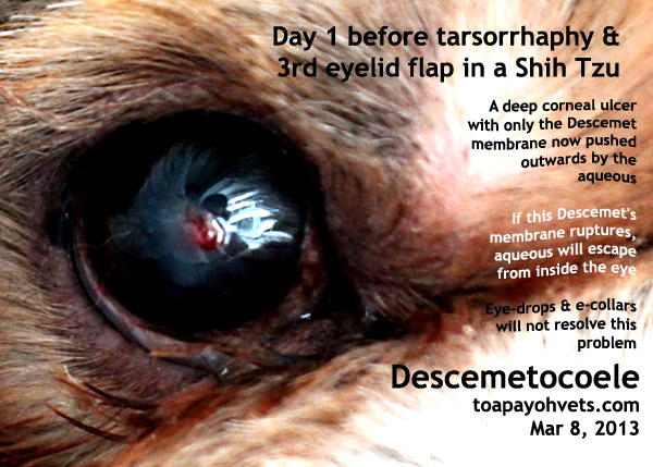
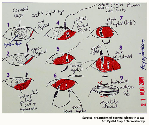
Veterinary And Travel Stories 3229 Intern Tarsorrhaphy And 3rd Eyelid Flap Procedures For Eye Corneal Ulcerations In Toa Payoh Vets
Dog Cat Ringworm Treatment Thornleigh Veterinary Hospital

Corneal Ulcers

Clinical Approach To The Canine Red Eye Today S Veterinary Practice

Full Text Traumatic Corneal Flap Displacement After Laser In Situ Keratomileusis Imcrj

The Concept Of Corneal Protection Clinician S Brief

Cornea Eye Diseases And Disorders Veterinary Manual
Non Healing Superficial Corneal Ulcers In Dogs Mspca Angell
Conjunctival Graft Technique In Dogs Vetlexicon Canis From Vetstream Definitive Veterinary Intelligence

A Modified Technique Of Keratoleptynsis Letter Box For Treatment Of Canine Corneal Edema Associated With Endothelial Dysfunction Giannikaki Veterinary Ophthalmology Wiley Online Library

Pdf Superficial Keratectomy And 360 Conjunctival Flap For Bullous Keratopathy In A Dog A Case Report
Surgery Of The Eye Veterian Key
Corneal Lipid
Understanding Canine Ocular Ulcers Dvm 360

Superficial Keratectomy And 360º Conjunctival Flap For Bullous Keratopathy In A Dog A Case Report
Toa Payoh Vets Singapore

Corneal Endothelial Dystrophy Dog Washington Dc Avo

How To Place A 3rd Eyelid Flap Into A Dog Or Cat Vetgirl Vet Ce Video
Http Www Eye Vet Com Wp Content Uploads 19 05 Diagnosis And Treatment Of Corneal Ulcerations Pdf
Third Eyelid Flap Technique In Dogs Vetlexicon Canis From Vetstream Definitive Veterinary Intelligence
Http Centredmv Com Wp Content Uploads 13 11 Fiche Ulcere Corneen Infecte An Pdf

3 Ways To Treat Canine Corneal Ulcers Wikihow

Treating A Corneal Ulcer In A Bug Eyed Dog Youtube

When Is It The Right Time To Operate On Indolent Ulcers Bsava12 Vin

The Golden Rules Of Corneal Ulcer Management Wsava13 Vin
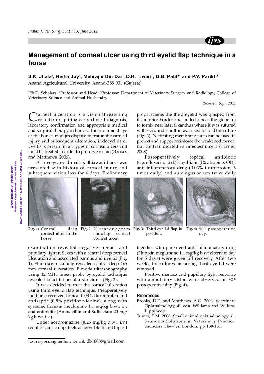
Pdf Management Of Corneal Ulcer Using Third Eyelid Flap Technique In A Horse

Corneal Ulcers In Dogs

Corneal Ulcers On Your Dogs Or Cat S Eye
Corneal Grafts At Animal Eye Care

My Dog Had A Third Eye Lid Flap Today Around 3 00pm I Picked Her Up St 5 00 She Has The Cone Collar That Is Really

Corneal Grafts Davies Veterinary Specialists

Conjunctival Pedicle Grafting Of The Cornea Information

Feline Corneal Sequestrum Mspca Angell
Corneal Disease Inherited In Dogs Petmd

Dry Eye Syndrome Wikipedia
Canine Dog Veterinary Surgery Anaesthesiaveterinary Surgery Anaesthesia Singapore Toa Payoh Vets Hamster Medicine Surgery Cases Health Sickness Singapore Singapore Toa Payoh Vets
Uc Davis Ophthalmologists Save Dog S Eyes Following Accident School Of Veterinary Medicine

Crsteurope Aggressive Recurrent Inflammation After Lasik
/Prolapsed_gland_of_the_third_eyelid-580285895f9b5805c23baa31.jpeg)
How To Treat Cherry Eye In Dogs
Surgery Of The Cornea And Sclera Veterian Key
1

Figure 2 From Key Facts Management Of Deep Corneal Ulcers Semantic Scholar

I Think My Dog S Scratched His Eye What Should I Do Goddard Veterinary Group
Corneal Ulcer Treatment Dog Eye Ulcer Treatment In Brisbane

How To Place A 3rd Eyelid Flap Into A Dog Or Cat Vetgirl Vet Ce Video

Experimental Lamellar Corneal Graft In Dogs Using Preserved Equine Pericardium
09asingapore Veterinary Dog Veterinary Tumours

Corneal Grafts Davies Veterinary Specialists
Why Do Dogs Have A Third Eyelid Healthy Dogs Animal Planet
Www Agriculturejournals Cz Publicfiles 139 17 Vetmed Pdf
Canine Dog Veterinary Surgery Anaesthesiaveterinary Surgery Anaesthesia Singapore Toa Payoh Vets Hamster Medicine Surgery Cases Health Sickness Singapore Singapore Toa Payoh Vets

The Concept Of Corneal Protection Clinician S Brief
Corneal Ulcer Treatment Dog Eye Ulcer Treatment Seah
Www Vettimes Co Uk App Uploads Wp Post To Pdf Enhanced Cache 1 Treating Corneal Ulceration In Dogs Part 2 Deep Ulcers Pdf

Observations In Ophthalmology Corneal Opacities In Dogs Cats

dental Scaling Health Care Problems In Singapore Dogs Fistula Oronasal Dog Toapayohvets Veterinary Vets Dog Cat Rabbits Hamster Veterinarian Veterinary Fees Services Education Stories Published By Toa Payoh Vets
Canine Dog Veterinary Surgery Anaesthesiaveterinary Surgery Anaesthesia Singapore Toa Payoh Vets Hamster Medicine Surgery Cases Health Sickness Singapore Singapore Toa Payoh Vets

3 Ways To Treat Canine Corneal Ulcers Wikihow
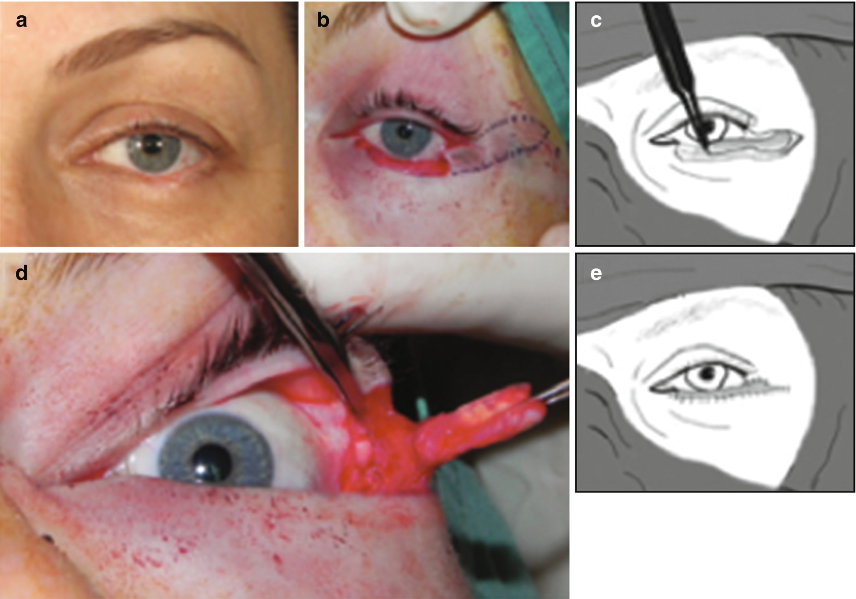
Principles And Techniques Of Eyelid Reconstruction Springerlink
Veterinary And Travel Stories March 13
Refractory Corneal Ulcer Management In Dogs Vet Med At Illinois

Eye Problems In Dogs And Cats Treatment Of Corneal Ulcers The Hi Lo

Corneal Grafts Davies Veterinary Specialists

The Concept Of Corneal Protection Clinician S Brief
Q Tbn And9gcse 042bwx Krcpoa2rh8unhs4bibxqnkab1unoimsohhmh0m Usqp Cau

Corneal Endothelial Dystrophy Dog Washington Dc Avo

Gundersen Flap In Severe Corneal Edema Youtube
Q Tbn And9gcsf6urf1qvyyg1bh32hzbldxegtaxugwzffnxqtxcr5dmkxaomr Usqp Cau

Deep Corneal Ulceration And Corneal Grafting Procedures
Pet Corneal Ulcers Animal Eye Consultants In Chicago Il

Deep Corneal Ulceration And Corneal Grafting Procedures
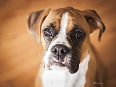
Eye Ulcers A Common Condition In Boxers And Other Adult Dogs


